How We Do It
Unlike other works of art, Little Cord Art pieces are custom-made using techniques applied in diagnostic medical pathology. Umbilical cord histology is different for every individual; therefore, no two Little Cord Art pieces will ever be the same. In addition, because we use a pathologic process to create our art, these images cannot be created via graphic design, painting, or other artistic methods. Both the subject and the art form are singular.
There are several labor-intensive steps involved in making Little Cord Art. Here’s how we do it (in a nutshell)…

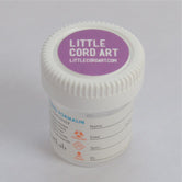
1 The umbilical cord specimen is collected by a doctor, nurse, medical assistant, or midwife at the time of baby’s birth and preserved in buffered formalin to “fix” the tissue. This preserves the cellular architecture, allowing us to capture the details that appear in the final art piece. Learn about our collection kit.

2 When we receive your cord sample, we set it in paraffin wax, slice individual specimens, and place them on microscope slides. Trained technicians process the cord in a state-of-the-art pathology lab using the latest equipment and techniques.

3 Next, we treat the cut specimens with various stains that make the cellular details visible to the naked eye. Each stain produces a unique color scheme. We choose which stains to apply based on the style(s) you select in the order process.

4 We produce 3-5 slides of varying color schemes and examine them under a high resolution light microscope that magnifies the cord tissue up to 400x the initial size.

5 We photograph the magnified images using a high-resolution camera mounted on the scope. We can zoom in on certain areas or crop the photos, creating unique images.

6 The resulting photos are printed with archival inks on high-quality canvas and stretched over a frame, producing a gallery-worthy piece of art.
